Stress Echocardiography is a specialised medical imaging technique to assess the heart’s function and blood flow under stress conditions. This diagnostic test combines echocardiography, which utilises ultrasound waves to create images of the heart, with physical stress to evaluate the heart’s response to increased workload.
Stress Echocardiography assesses your heart function under stress and can show problems that aren’t visible when your heart is resting. It provides valuable information about cardiac function, identifies coronary artery disease, and helps healthcare professionals assess the heart’s performance during exertion or induced stress.
What Is Stress Echocardiography?
Stress Echocardiography is a diagnostic procedure that involves creating detailed images of the heart before and after inducing stress on the cardiovascular system. The echocardiogram, performed at rest and during stress, allows healthcare professionals to evaluate changes in the heart’s contraction and blood flow during stress.
This test is beneficial for detecting coronary artery disease, assessing heart valve function, identifying areas of the heart with compromised blood supply, and provides critical information for diagnosis and treatment planning.
Who Needs Stress Echocardiography?
Stress echocardiography is most commonly used in the diagnosis of significant coronary artery disease. This condition occurs when the blood vessels that carry blood to your heart muscle become blocked. When this happens, the heart muscle that receives blood from that vessel may not function well under stress. It can also be used to monitor valvular heart disease and assess how well your heart relaxes when under stress. It can be useful to assess symptoms of chest pain, breathlessness and dizziness or fainting episodes particularly when related to exertion. Other people who may undergo an exercise stress echocardiogram would include screening for athletes or patients prior to undergoing surgery.
How Do I Prepare For Stress Echocardiography?
Your doctor will provide guidance on medication management, including any temporary withholdings if needed. Wear comfortable clothing and shoes that you can exercise in.
What Happens During Stress Echocardiography?
The following process will take place when you come for the test:
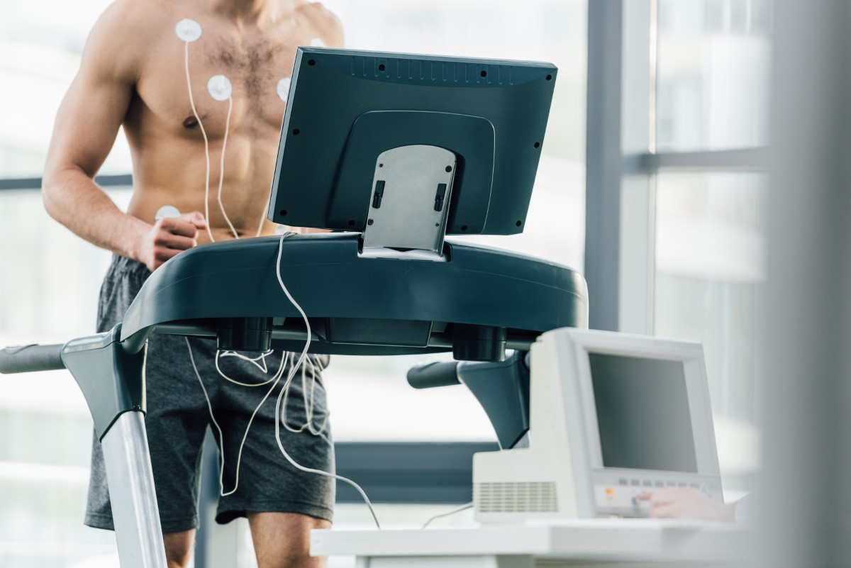
- Small electrodes will be placed on your chest to allow monitoring of your heart rate. You will also wear a blood pressure cuff to record your blood pressure during the test.
- You will be asked to lie on your left side to allow the sonographer to acquire your baseline images. Gel will be applied to your chest, and a probe will be placed at various positions on your chest to acquire images of your heart at rest.
- You will then be asked to exercise on a treadmill, starting slowly then gradually increasing in intensity every 3 minutes. Throughout this phase, continuous monitoring of vital signs and ECG ensures safety. When you have reached the limit of your exercise tolerance the treadmill will be stopped and you will be asked to return to the exam table for additional images of your heart to be recorded.
- After the final echocardiogram you will be monitored until your heart rate and blood pressure returns to normal before you return home.
The test takes about 30 minutes to 1 hour to complete.
What To Do After Stress Echocardiography?
You will be able to resume your usual daily activites after the test. The test results will be reviewed by your Cardiologist, and follow-up will be arranged to discuss the findings and treatment plan for you.
What are the Risks of undergoing a Stress Echocardiogram?
Exercise stress echocardioraphy is a safe test with minimal side effects. Serious complications are extremely rare and come with stressing the heart which can lead to abnormal heart rhythms or chest pain particularly in the presence of underlying heart disease. Additional risk may come from injury falling from the treadmill machine. You will be closely monitored for side effects during and after the test.


